
The solution
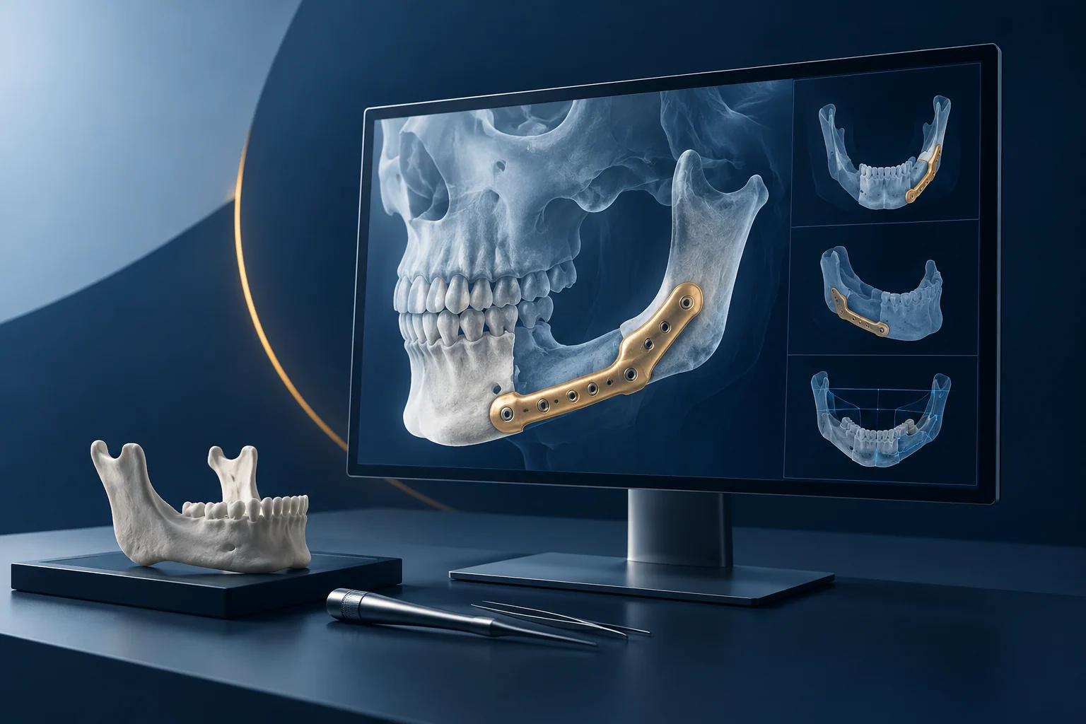
A specialist medical software application for visualizing anatomical data and supporting mandible reconstruction and surgical planning. The platform combines medical-image import and processing, anatomical segmentation, 3D mandible and reconstruction geometry visualization, and interactive inspection tools in a professional workstation experience. It also covers mesh, volume, and anatomical-structure manipulation, reusable planning and inspection tools, and optimized model inference intended to execute imaging models efficiently inside the application.
The challenge
Healthcare and life-sciences organizations often manage complex imaging and surgical-planning work across multiple tools. For mandible reconstruction, specialists need to review anatomical data, understand bone and reconstruction geometry, and prepare a plan that can be inspected and discussed. When image import, segmentation, 3D visualization, and planning tools sit in separate environments, teams may spend time moving data, reconciling views, and rebuilding context. The platform addresses that friction by describing a more integrated workstation for medical-image processing, anatomical segmentation, and 3D mandible and reconstruction geometry visualization.
Core capabilities
- Medical image import and processing: bringing imaging data into the application for review and downstream planning steps.
- Anatomical segmentation: separating relevant anatomical structures to support focused inspection and 3D visualization.
- 3D mandible and reconstruction geometry visualization: viewing mandible anatomy and reconstruction geometry in three dimensions.
- Interactive inspection and surgical-planning workflows: tools for examining anatomy and preparing planning views.
- Mesh, volume, and anatomical-structure manipulation: working with different representations of clinical data.
- Optimized model inference: inference components designed to execute imaging models efficiently within the application.
- Reusable planning and inspection tools: repeatable tools supporting consistent preparation and review.
How it works
The software workflow follows these stages:
- Import: medical images are brought into the workstation for processing.
- Process and segment: imaging data is prepared and anatomical structures are segmented to isolate relevant regions.
- Visualize: the mandible and reconstruction geometry are reviewed in 3D.
- Inspect and manipulate: mesh, volume, and anatomical structures are examined and adjusted using interactive tools.
- Plan: reusable planning and inspection tools support the preparation of a surgical-planning view.
- Run inference: optimized inference components execute imaging models within the application to support responsive analysis and visualization.
Business value
The platform brings image processing, anatomy visualization and reconstruction planning into a connected specialist workflow. It gives clinical and technical teams a consistent environment for examining geometry, reviewing cases and coordinating preparation.